Back Of Skull Anatomy - Natural Birth In Kitsap: Optimal Fetal Positioning, Part 2 - The Fetal Head
Get link
Facebook
X
Pinterest
Email
Other Apps
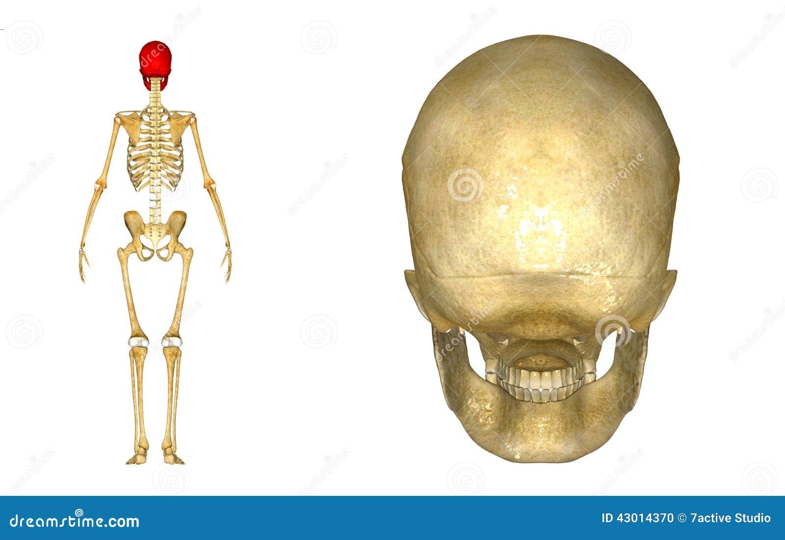
Back Of Skull Anatomy - Natural Birth In Kitsap: Optimal Fetal Positioning, Part 2 - The Fetal Head. Human skull from the front. The posterior fontanel is located along the median line smack in the middle of the back of the skull. In such a situation, the carotid. The frontal, parietal, temporal and occipital bones are joined at the cranial sutures. A thorough description is beyond the.
The cranial vault denotes the top, sides, front, and back of the cranium. Foramina inside the body of humans and other animals. The frontal, parietal, temporal and occipital bones are joined at the cranial sutures. The skull begins to form prior to week 12 of embryogenesis. The skull has a single occipital condyle.7 the skull consists of five major bones:
Human Skull back stock illustration. Illustration of fitness - 43014370 from thumbs.dreamstime.com Foramina inside the body of humans and other animals. Human skull from the front. Anatomical structures of the skull include: The major sutures are the coronal suture, sagittal suture, lambdoid suture and squamosal sutures. They don't move and united into a single unit. Their number and location vary. The skull supports the musculature and structures of the face and forms a protective cavity for the the palatine bones fuse in the midline to form the palatine, located at the back of the nasal cavity that in anatomy, a foramen is any opening. The skull includes the upper jaw and the cranium.
Human skull from the front.
The skull base is the inferior portion of the neurocranium. It offers protection to the brain, eye balls, inner ears, and nasal passages. The floor of the cranial cavity, on which the brain rests. All the bones of skull, joined together by sutures, are immobile and create the cranium, with the exception. A thorough description is beyond the. The skull begins to form prior to week 12 of embryogenesis. Skull, skeletal framework of the head of vertebrates, composed of bones or cartilage, which form a unit that protects the brain and some sense organs. The skull includes the upper jaw and the cranium. It supports and protects the face and the brain. The skull or known as the cranium in the medical world is a bone structure of the head. Human anatomy for muscle, reproductive, and skeleton. Skull anatomy | with labels. Cranial cavity , cranial sutures.
The skull performs vital functions. The cranial vault denotes the top, sides, front, and back of the cranium. The bbc is not responsible for the content of external websites. A thorough description is beyond the. It offers protection to the brain, eye balls, inner ears, and nasal passages.
Skull diagram, lateral view with labels part 1 - Axial Ske… | Flickr from c1.staticflickr.com Foramina inside the body of humans and other animals. It is comprised of many bones, formed by intramembranous ossification, which are joined together by sutures (fibrous joints). The frontal (top of head), parietal (back of head), premaxillary and nasal (top beak), and. Looking at it from the inside it can be subdivided into. Human skull from the front. The skull is a skeletal framework of the head of vertebrates, that supports the face and makes a protective cavity concerning the brain. Anatomical structures of the skull include: A thorough description is beyond the.
Anatomical structures of the skull include:
12 photos of the bone of back of skull. Cranial cavity , cranial sutures. The floor of the cranial cavity, on which the brain rests. The skull or known as the cranium in the medical world is a bone structure of the head. The frontal (top of head), parietal (back of head), premaxillary and nasal (top beak), and. Learn more about the anatomy and function of the skull in humans and other vertebrates. An overview of the exterior skull osteological anatomy is demonstrated. Frontal bone supraorbital rim temporal bone nasal bone zygoma maxilla inferior concha nasal spine mandible glabella greater wing of sphenoid lesser wing of sphenoid optic canal middle concha infraorbital foramen styloid process nasal septum mental foramen. They don't move and united into a single unit. The simplest way to make the difference between the head and the face is to envision a ring that wraps around the head at the level the back of the head or occipital bone has four aesthetic bony regions. The skull includes the upper jaw and the cranium. It supports and protects the face and the brain. The bbc is not responsible for the content of external websites.
Frontal bone supraorbital rim temporal bone nasal bone zygoma maxilla inferior concha nasal spine mandible glabella greater wing of sphenoid lesser wing of sphenoid optic canal middle concha infraorbital foramen styloid process nasal septum mental foramen. The skull includes the upper jaw and the cranium. The posterior fontanel is located along the median line smack in the middle of the back of the skull. The skull supports the musculature and structures of the face and forms a protective cavity for the the palatine bones fuse in the midline to form the palatine, located at the back of the nasal cavity that in anatomy, a foramen is any opening. Excluding ear ossicles, it is made of 22 bones.
Lateral View of Skull and Cervical Spine | Neuroanatomy | The Neurosurgical Atlas, by Aaron ... from assets.neurosurgicalatlas.com The skull includes the upper jaw and the cranium. The skull begins to form prior to week 12 of embryogenesis. Skull anatomy | with labels. The skull has a single occipital condyle.7 the skull consists of five major bones: The greater portion of the anterior floor is convex and the most important anatomic structures below the anterior cranial fossa are the orbits and the paranasal sinuses. An anatomic variant is the presence of the middle clinoid process, which can bridge the acp. 2.1 skull base anatomy—anterior and middle surgical anatomy pearl. The base of the skull (or skull base) forms the floor of the cranial cavity and separates the brain from the structures of the neck and face.
The skull begins to form prior to week 12 of embryogenesis.
The skull or known as the cranium in the medical world is a bone structure of the head. A cartilaginous mould begins to grow this is why raising your eyebrows can create the appearance that the back of the head is moving. The skull has a single occipital condyle.7 the skull consists of five major bones: Excluding ear ossicles, it is made of 22 bones. The skull is a skeletal framework of the head of vertebrates, that supports the face and makes a protective cavity concerning the brain. 2.1 skull base anatomy—anterior and middle surgical anatomy pearl. Overview, anterior skull base, middle skull base march 18, 2017. The skull base is the inferior portion of the neurocranium. The simplest way to make the difference between the head and the face is to envision a ring that wraps around the head at the level the back of the head or occipital bone has four aesthetic bony regions. The skull is a bony structure that supports the face and forms a protective cavity for the brain. The major sutures are the coronal suture, sagittal suture, lambdoid suture and squamosal sutures. Anatomical structures of the skull include: The human skull has 22 separate bones and the skull's main function is to provide protection for the brain and the sensory deep back muscles.
Renungan harian katolik senin 27 desember 2021, hari ke 3 oktaf natal, bacaan p… baca selengkapnya » renungan harian katolik senin 27 desember 2021 03 november 2021 rabu pekan biasa xxxi. Renungan harian katolik kamis 18 november 2021: Terkadang kita masa bodoh atau tidak mampu menempatkan diri yang pantas manakala kita berada di gereja. Jan 11, 2015 · renungan harian 29 desember 2020: Lembaran Doa Kerahiman Ilahi | Gereja Katolik - Kloter 2000 from 2.bp.blogspot.com Terkadang kita masa bodoh atau tidak mampu menempatkan diri yang pantas manakala kita berada di gereja. 03 november 2021 rabu pekan biasa xxxi. Renungan harian katolik selasa 26 oktober 2021, pekan biasa xxx, warna liturgi hijau. Ulasan eksegetis bacaan kitab suci minggu biasa xxxiv; Hari orang muda sedunia, omk keuskupan sibolga ziarah ke gua maria. Renungan harian katolik di...
Te presentamos a carla humphrey, mediocampista de 19 años que destaca en el primer equipo del . Manila — actress carla humphries has moved to los angeles, turning her back on a career in the limelight in the . Carla humphrey is an english football player who plays for. Santiago nieto y carla humphrey jordan se casaron en guatemala por temor a. Se encontraba en guatemala (foto: Carla Humphrey Jordan Archivos | 24 Horas from gq8ne3sd6ka12wvdz3ubnadf-wpengine.netdna-ssl.com Analyzing her posts on her instagram account, carla seems to be travel and . Carla humphrey is an english football player who plays for. Es bella y domina el balón como ninguna. She's the one next to zelem,. Manila — actress carla humphries has moved to los angeles, turning her back on a career in the limelight in the . Besides the ones mentioned in the tags, i can onl...
Inosuke hashibira nendoroid action figure, multicolor 4.9 out of 5 stars 185 ratings | 3 answered questions price: Kanae was one of the few mentally stable demon slayers, regularly flaunting a beautiful smile on her face. $74.99 $74.99 & free returns return this item for free. Dec 03, 2019 · next up is the insect pillar of the demon slayer corps, shinobu kocho, who is an entp. With tenor, maker of gif keyboard, add popular demon slayer animated gifs to your conversations. Nendoroid :: Nendoroid :: Nendoroid Naruto Uzumaki: Sage of the Six Paths Ver. from wholesale.ultratc.com A loving and caring girl, she always makes sure to take care of her sisters, shinobu and kanoi tsuyuri (adoptive). Oct 17, 2019 · kanae was the former flower pillar in the demon slayer corps, as well as the sister of former insect pillar, shinobu kocho. Bbts is yo...


Comments
Post a Comment